Spotlight on amazing Australian research
Interview with Linda Rogers, Chris O’Brien Lifehouse Comprehensive Cancer Hospital, Sydney, Australia.
Highlighted research tool: GelCount™ by Oxford Optronix.
Linda Rogers is currently the PC2 laboratory manager in the Arto Hardy Biomedical Innovation Hub at Chris O’Brien Lifehouse, a comprehensive cancer hospital located in Camperdown, Sydney, Australia.
At the time of undertaking the research presently highlighted, Linda was Radiobiology Laboratory Manager at Chris O’Brien Lifehouse. Along with her collaborators, A/Prof Natalka Suchowerska and Prof David McKenzie, Linda began this research project as an avid user of our first-generation colony counting system, the ColCount, followed by our much more capable GelCount system. Since Mrs. Rogers has years of experience in cell biology and assay development, we were thrilled when Linda agreed to contribute to our ongoing customer spotlight and talk about her oncology research goals and newest publication in the International Journal of Radiation Biology.
Can you describe your research?
We took a new approach to kill cancer more effectively using a striped radiation beam pattern and compared the results with those from a conventional uniform beam. We also split the treatment into two equal parts, separated by a time of between minutes and hours. We measured the success of the treatment by looking at whether surviving cancer cells are capable of dividing enough times to form a colony of cells, leading to a new tumour. We know that some cells survive radiation treatment, but only divide a couple of times and then stay inactive without reforming the cancer.
What are youR main findings to date?
We found that for cancer cells, splitting the treatment into two deliveries of half the dose each time, results in more cancer cell killing, than delivering the same dose in a single treatment, however this was not seen in the normal cells. This fascinating observation gives us the option of either giving the SAME dose to get more cancer cell death or LESS dose for the same effect. For prostate cancer we get up to 17% more cancer killing when we split the dose delivery.
On top of this split dose effect, using the striped beam pattern resulted in even more killing of cancer cells, for example, in lung cancer, we found a further 4.6% increase in cancer reduction. Furthermore, when we looked at the therapeutic ratio (defined as the ratio of survival of the normal prostate relative to prostate cancer), we found an increase in this ratio of up to 27% by both splitting the dose and using the striped radiation beam. A high therapeutic ratio means more cancer cells are killed for a given treatment, while the normal cells survive better, which is exactly what we want inside the human body.
We believe the cause for our findings is related to the bystander effect, whereby irradiated cells send out signals to neighbouring unirradiated cells, affecting their survival. Please see figure 4 in our paper, entitled: “Radiation Responses of Cancer and Normal Cells to Split Dose Fractions with Uniform and Grid fields: Increasing the Therapeutic Ratio” published online in the International Journal of Radiation Biology.
These bystander signalling factors, released by cells after the first dose, communicate and “prime” the cells for the second dose, making them more vulnerable and increasing cancer cell killing.
Can you summarize your new paper and the main findings?
The paper, published online this year in the International Journal of Radiation Biology, is entitled “Radiation Responses of Cancer and Normal Cells to Split Dose Fractions with Uniform and Grid fields: Increasing the Therapeutic Ratio” (2022).
This is an in vitro study using cell lines including two prostate cancer, a normal prostate, a non-small cell lung cancer, a tongue cancer and a glioma. We examined the effects of both temporal and spatial modulation of radiation beams on in vitro cell survival. The paper summarises the findings that we describe above.
How do you use GelCount by Oxford Optronix?
We often look at whether any surviving cancer cells would be capable of dividing enough times to form a colony of cells and then possibly a whole new tumour. Some cells may survive but only divide a couple of times and then stay inactive without reforming the cancer. This approach, called a clonogenic assay, specifically identifies how many cancer colonies form after treatment. The Oxford Optronix GelCount™ has enabled us to perform the clonogenic assay by characterising each cell type and parameterising their specific behaviour and appearance. In this way we were able to efficiently and reliably identify colonies defined as those with more than 50 cells (or 6 cell divisions). We studied two different prostate cancer cell lines (hormone sensitive and hormone insensitive), a normal prostate cell line, a lung cancer (non small-cell lung) and brain cancer (glioma).
Why is GelCount helpful?
The GelCount™ is a greatly improved version over its older version, the ColCount. The old ColCount only examined one flask at a time. To count one experiment, which could be up to 100 flasks, this would be very time laborious. The old ColCount also could not scan and count multi-well plates. The GelCount not only can accurately scan and count multi-well plates, but its resolution is also far improved and can count up to four multi-well plates at a time or 8 x T25cm2 flasks at a time.
There is also opportunity for customising the instrument for specific needs. Several trays are available with the equipment that fit only certain brand of flasks. APAC Scientific (Australia), along with Oxford Optronix, have an amazing technical support team and custom-built a new tray for our purposes. There were also some areas within the program where we needed parameters customised and the amazing team wrote some code into the program to meet our needs.
Since the clonogenic assay is the gold standard assay used in in vitro radiobiology studies, the GelCount™ will be a timeless addition to any radiobiology laboratory.
What are your future research goals?
We would love to translate these findings into the clinic. The good aspect from the clinical point of view is that we don’t need to change any of the equipment, as our modern radiation treatment facilities already allow for striped beams and splitting the dose into two parts only fifteen to forty minutes apart.
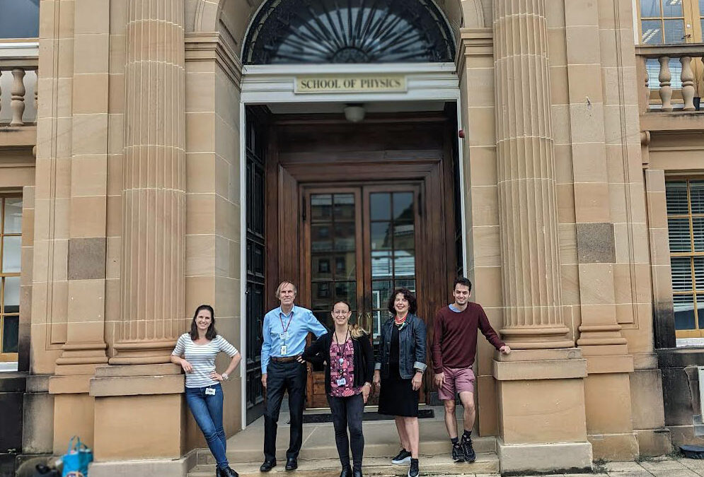
The Research Team
 From left to right: Juliette Harley, David McKenzie, Linda Rogers, Natalka Suchowerska, Georgio Katsifis.
Linda Rogers is PC2 Laboratory Manager in the Arto Hardy Biomedical Innovation Hub at Chris O’Brien Lifehouse and is an expert in cell biology, especially cell survival assays.
Juliette Harley is a PhD student at the School of Physics, University of Sydney who is studying how to improve cancer treatments using radiation with novel therapies.
Prof David McKenzie is a professor of materials physics and is expert in applying physical science to medicine and biology.
A/Prof Natalka Suchowerska is a medical physicist and an expert in clinical applications of radiation treatments.

