
High-Frequency Ultrasound for Dermatology Research and Preclinical Imaging

See Deeper. Understand Skin Better.
SkinScanner™ is a dermoscopy-guided high-frequency ultrasound system designed for dermatology research and preclinical studies.
The device combines dermoscopy (optical skin imaging) with high-frequency ultrasound (20–40 MHz) to visualize skin structures below the surface while maintaining precise spatial alignment with visible skin features.
This enables researchers to perform non-invasive imaging of skin layers, lesions, and vascular structures, supporting quantitative dermatology research and longitudinal studies.
MDR (CE) certified.
SKINSCANNER

SkinScanner Handheld Dermatology Device
It integrates:
-
Optical dermoscopy guidance
-
High-frequency ultrasound imaging
-
Research software for data analysis
This combination allows researchers to correlate surface skin features with subsurface tissue structure, which is difficult with most existing dermatology imaging technologies.
Key Technical Specifications
Imaging Technology
-
High-frequency ultrasound: 20–40 MHz
-
Maximum imaging depth: ≤10 mm
-
Optical dermoscopy field of view: 12 × 12 mm
System Features
-
Dermoscopy-guided ultrasound positioning
-
High-resolution and high-depth imaging modes
-
Positioning line for intuitive image interpretation
-
Reproducible scan positioning
-
Portable handheld system
These capabilities allow researchers to capture consistent skin imaging data across multiple scans and time points.
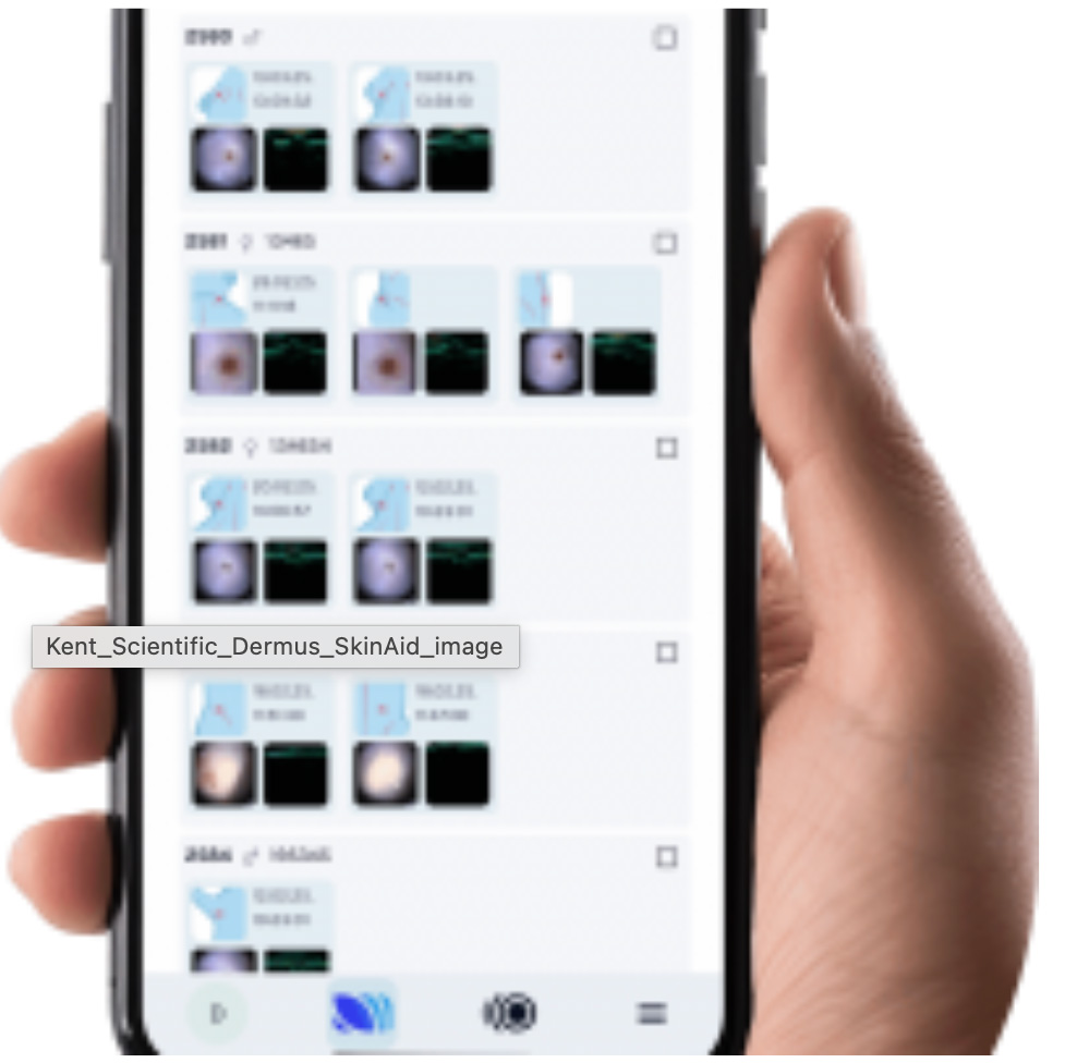
SkinAid™ Research Software Platform
SkinScanner includes the SkinAid™ research software package, designed to support preclinical data collection and dermatologic imaging analysis.
 Intelligent Skin Imaging
-
AI-powered skin layer segmentation for precise analysis
-
Quantified skin change tracking for objective insights
-
Secure, anonymized patient data management
-
Web-based platform with cloud or local storage
-
Advanced filters, settings, and annotation tools
-
Seamless third-party image import
-
Two-point distance and volume measurement for accurate assessment
Typical Applications
-
Skin cancer research (melanoma, basal cell carcinoma, squamous cell carcinoma)
-
Inflammatory skin diseases such as psoriasis and morphea
-
Hidradenitis suppurativa research
-
Hair follicle and scalp imaging
- Cosmetic and dermatologic treatment studies
HUMAN-USE & Animal Models
As well as human-use, the high-frequency skin ultrasound suits translational studies on rodent models and mini pigs because their skin thickness fits well within the ≤10 mm imaging depth.

Frequently Asked Questions
What is dermoscopy-guided high-frequency ultrasound?
Dermoscopy-guided high-frequency ultrasound is an imaging approach that integrates optical dermoscopy with high-frequency ultrasound imaging (20–40 MHz) to enable spatially aligned visualization of surface morphology and subsurface skin structures.
This combined modality allows researchers to correlate dermoscopic features with ultrasound-derived information on skin layer architecture, lesion morphology, and vascular characteristics.
What is SkinScanner designed for?
SkinScanner is a handheld high-frequency ultrasound system developed for dermatology research and preclinical imaging studies. It is also approved for clinical use in European countries.
The system enables non-invasive imaging of superficial tissues and supports applications including:
-
structural analysis of skin layers
-
imaging of benign and malignant skin lesions
-
evaluation of inflammatory skin disease models
-
monitoring of structural changes in longitudinal studies
-
imaging in experimental dermatology and preclinical models
What ultrasound frequencies are used for dermatologic imaging?
Dermatologic ultrasound typically uses high-frequency transducers in the range of 20–50 MHz, which provide high spatial resolution for imaging superficial tissues.
SkinScanner operates at 20–40 MHz, enabling high-resolution imaging of skin structures with a penetration depth of approximately 10 mm.
What tissue depth can be visualized using high-frequency ultrasound?
High-frequency ultrasound used in dermatologic imaging typically visualizes tissue structures within the epidermis, dermis, and superficial subcutaneous tissue, generally up to depths of approximately 5–10 mm.
This depth range is appropriate for imaging most dermatologic lesions and structural skin changes.
What advantages does dermoscopy-guided ultrasound provide?
Dermoscopy-guided ultrasound allows precise spatial alignment between optical surface imaging and ultrasound acquisition.
This improves:
-
scan positioning accuracy
-
reproducibility in longitudinal imaging studies
-
correlation between dermoscopic morphology and subsurface tissue structure
Such alignment can be valuable in research settings requiring repeated imaging of defined skin regions.
What dermatology research applications use high-frequency ultrasound?
High-frequency ultrasound is used in multiple areas of dermatologic research, including:
-
skin cancer research (melanoma, basal cell carcinoma, squamous cell carcinoma)
-
inflammatory skin diseases such as psoriasis and morphea
-
hidradenitis suppurativa research
-
hair follicle and scalp imaging
-
cosmetic and dermatologic treatment studies
It is also widely used in experimental dermatology and translational research.
What peer-reviewed studies have investigated dermoscopy-guided high-frequency ultrasound?
Dermoscopy-guided high-frequency ultrasound has been investigated in several peer-reviewed studies examining dermatologic imaging and preclinical skin models.
Recent publications include:
-
Dermoscopy-guided high-frequency ultrasound for preoperative assessment of basal cell carcinoma lateral margins: A pilot study — British Journal of Dermatology, 2025
-
Optically guided high-frequency ultrasound for estimation of Breslow thickness compared with multispectral imaging — Cancers, 2024
These studies illustrate the application of high-frequency ultrasound for tumor characterization, structural skin imaging, and experimental dermatology research.
Is high-frequency ultrasound suitable for preclinical dermatology studies?
Yes. High-frequency ultrasound is commonly used in preclinical dermatology research, including studies involving animal models of skin disease.
Because ultrasound imaging is non-invasive and repeatable, it enables longitudinal assessment of structural changes within the same tissue region over time.
Does dermoscopy guidance improve reproducibility in ultrasound imaging?
Dermoscopy guidance facilitates consistent positioning of the ultrasound scan relative to surface landmarks, which can improve reproducibility in repeated imaging sessions.
This can be particularly useful in longitudinal studies where imaging must be performed at the same anatomical location across multiple time points.